


Monitoring Disease Progression
PET/CT images of a mouse lungs after intranasal administration of an aerosol containing FDG. Intranasal administration is one of the main routes of allergen challenge in mouse models of airway disease. Although it is widely used, it is not well established the amount of allergen that reaches the lung or is lost to the gastrointestinal tract. The local distribution and the dynamics of the challenge solution within the airways were visualized using dynamic PET imaging co-registered with CT.
Mol Imaging Biol. 2009 Jul-Aug;11(4):263-8. doi: 10.1007/s11307-009-0199-y


(Left) CT images of a fish model are used to quantify size and density of different organs and masses under study. Scans are taken in a few minutes, and low dose protocols are used to minimize the radiation exposure of the sample.
Unpublished data.
(Bottom)
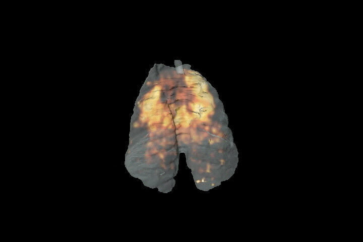
Left: Surface render of a mice lung study; low-dose CT imaging is used for the quantification of tissue densities in longitudinal protocols. Right: FDG image of a pulmonary infection reveals the increased metabolism of the affected tissue. These techniques are being used in oncology, COPD and infectious diseases.
Unpublished data.

Applications in cardiology
Semi-quantitative, static PET imaging is used to assess [13N]-ammonia myocardium perfusion. These studies are aimed to elucidating the effect of injecting human embryonic stem cell-derived (hES) hemangioblasts on infarcted rat hearts. NH3 myocardium perfusion image of a control animal (left) compared to a sick one (right).
Mol Imaging Biol. 2012 Oct;14(5):541-5. doi: 10.1007/s11307-011-0538-7
